

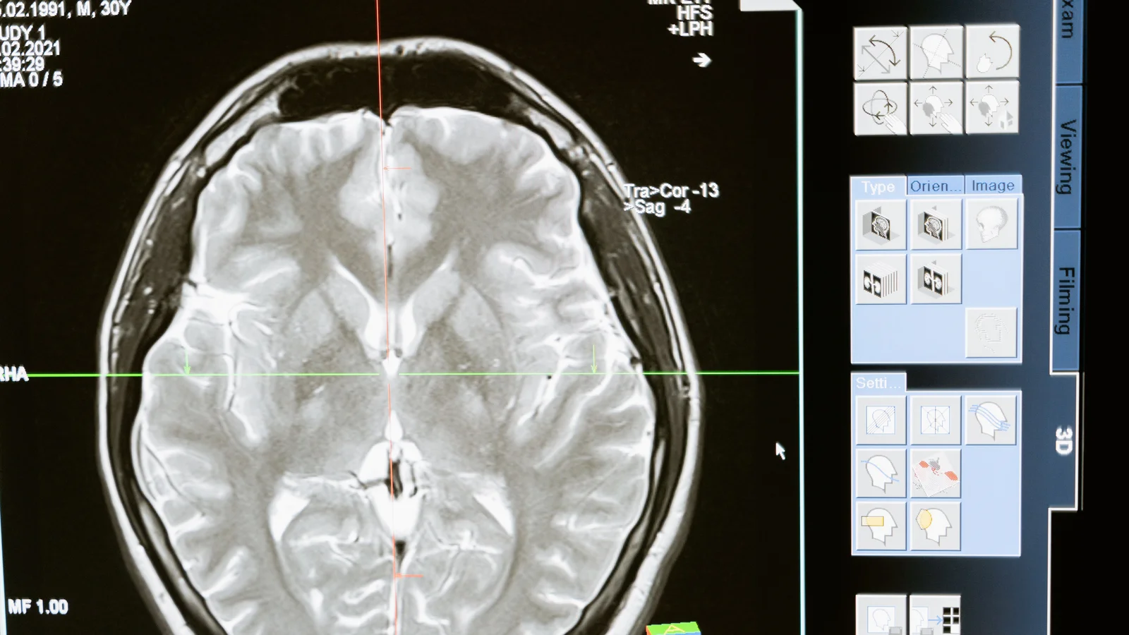
A fetus does not hold still for a scanner, and that simple fact has long frustrated prenatal imaging. The new model adjusts scans on the fly to follow fetal movement, MIT News reported, potentially sparing families return trips to the hospital. MRI has become a complement to ultrasound because it offers sharper soft-tissue contrast without ionizing radiation, yet motion during a slow acquisition degrades images and often forces a repeat exam, a review in the American Journal of Neuroradiology noted, with emotional costs for parents and budget strain for providers.
The approach hinges on a quick, low-resolution "navigator" image captured just before each diagnostic slice. From that rough snapshot, the network estimates the head's position and orientation, and the next slice is prescribed accordingly. The method, called E(3)-Pose, bakes two physical facts into the network's design, the researchers wrote in their paper: rotating the input rotates the answer in step, and the fetal head looks nearly identical when flipped left to right. That mirror symmetry is exactly what trips up conventional models, because underdeveloped anatomy and scan artifacts can hide features such as the eyes that would otherwise settle which way the head faces. Earlier automated systems estimated pose once at the start of a sequence and planned every slice from it, according to the paper, leaving motion between slices unaddressed.
Clinical use is the stated goal, though the work remains a research result. The authors report competitive accuracy on public research-grade datasets and state-of-the-art performance on clinically representative volumes, the paper states, and have released their code publicly. The project drew support from the MIT Jameel Clinic, the MIT-Takeda Program and the National Institutes of Health, with ethics approval from Boston Children's Hospital's review board. The researchers also suggest the symmetry-aware design could transfer to other localization problems involving noisy sensors and scarce training data.